
Patient – Questions For My Doctor
Clinical Breast Exam: Women in their 20s and 30s should have a clinical breast exam preferably every 3 years. After age 40, women should have a breast exam by a health expert every year. It’s a good idea to do a breast self-exam (BSE) before the clinical exam to become familiar with what your breast feel like.
Breast Awareness and Breast Self-Exam (BSE): BSE is an option for women starting in their 20s. If you do BSE on a regular basis, you will become familiar with how your own breasts feel and with this awareness, you are more likely to find a lump or suspicious area in the breast. The American Cancer Society has special programs to teach you how to examine your breast properly and cards describing the process can help you as you begin to examine yourself.
A technologist (usually a woman) will position you correctly for the exam and take the pictures.
- FNA: (Fine Needle Aspiration) In this test fluid is drawn out to ck the cells in the fluid for cancer. Your doctor may use ultrasound to guide him.
- Stereotactic core needle biopsy: In this test the needle is larger and removes several cylinders of tissue. The area is numbed and this test is usually done on an outpatient basis.
- Surgical biopsy: Sometimes surgery is needed to remove all or part of a lump so it can be looked at under the microscope. Usually the surgeon will remove the lump and some normal tissue around it. Usually this is done in the hospital on an outpatient basis. Occasionally, light sedation is used to make the patient more comfortable and less aware of the process. After the lump is removed, the lab will determine if it is cancer and then, if so, if it is invasive or not. The most common type of cancer (DCIS) is contained in the ducts and the cure rate is very good. The sample will also be graded ER-positive or PR-positive. This has to do with how the cancer responds to hormone treatment and enables your oncologist (cancer doctor) to determine the best treatment for you if the biopsy shows cancer.
Additional questions you could ask might include the following:
- Would you please write down the type of cancer I have?
- May I have a copy of my pathology report?
- Has the cancer spread to lymph nodes or other organs?
- What stage is my cancer? What does that mean?
- What treatment choices do I have? What do you recommend and why?
- Am I eligible for clinical trials?
- Will I lose my hair? If so, what can I do about it?
- How long will each treatment last? What can I expect?
- Can I drive myself home after the treatment or will I need help?
- What are the chances of my cancer coming back after the treatment?
- Should I follow a special diet?
- What kind of breast reconstruction is possible in my case?
- Will the treatment cause menopause?
- Can I still have children?
- What should I do to get ready for my treatment?
- Will the treatment affect breast sensation?
- What are my chances of survival based on the type of cancer I have?
- Ask NOAH about Cancer www.noah-health.org
- Association of Cancer Online Resources www.acor.org
- Breast Cancer Answers Project www.canceranswers.org
- National Breast Cancer Coalition www.natlbcc.org
CTLM® – Laser Breast
Imaging Without Compression
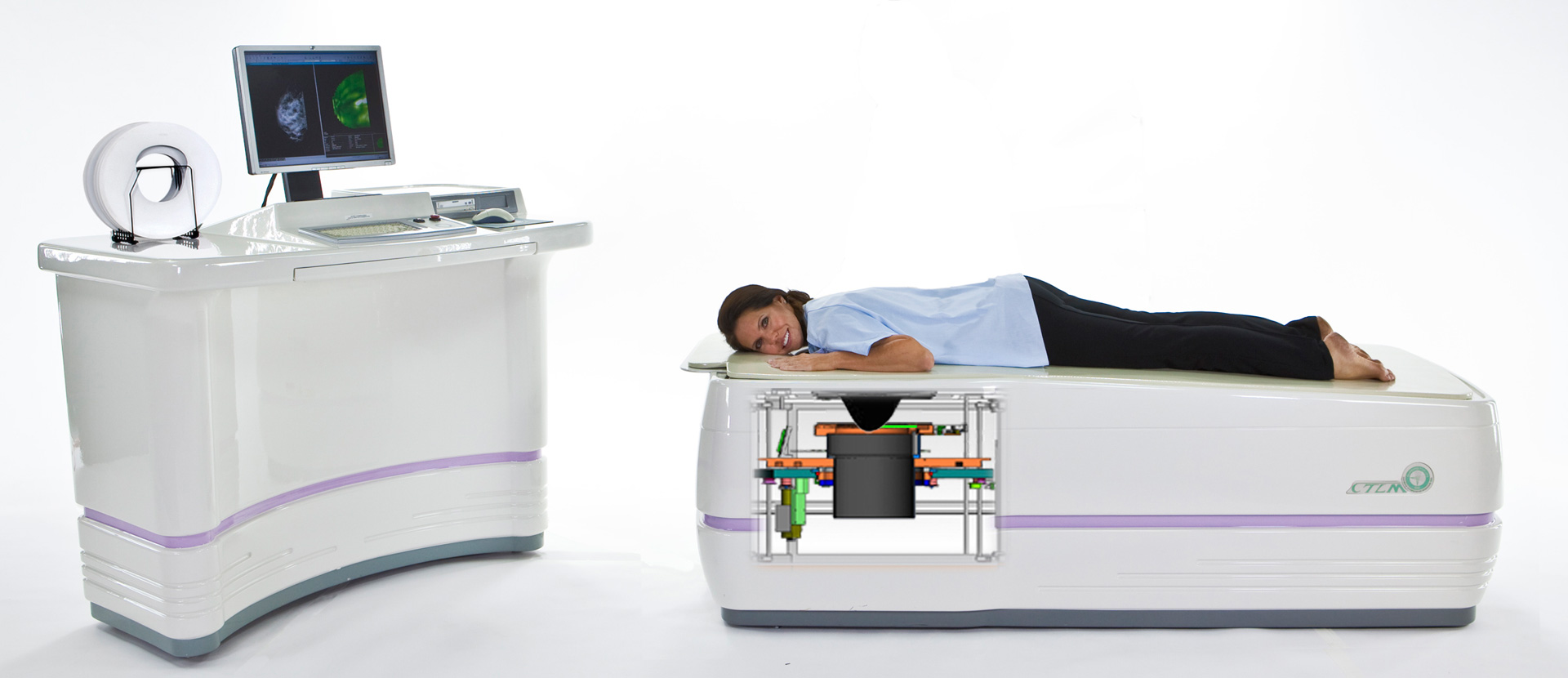
Facts About CTLM® And Optical Imaging:
- CTLM® – Computed Tomography Laser Mammography is part of the emerging field of optical imaging.
- Scientific data has demonstrated that CTLM® images angiogenesis. Angiogenesis is a requirement for any process in the breast that has increased metabolic demands such as invasive and in situ cancers.
- CTLM® does not use ionizing radiation (no x-rays).
- CTLM® was designed as an adjunct to mammography and ultrasound for imaging dense breasts.
- There is NO breast compression with CTLM® and the breast hangs in the machine opening in it’s natural position.
- In a study of over 100 women, including 30 with breast cancer, optical imaging increased sensitivity and specificity of breast cancer detection by more than 90% (Britton Chance, Molecular Imaging, Vol. 2 #2)
- CTLM® – Computed Tomography Laser Mammography provides additional biological information that may be useful in assisting with the early detection of breast cancer and/or clinically relevant anomalies.